

The famous Italian anatomist Gabriel Fallopian in the mid-1500s first described the appearance of what later came to be called the fallopian tubes.
These two passageways connect the uterus with the area around the ovaries.
they are 3 to 5 inches (7.5 to 12.5 cm) in length and very narrow and fragile.
The opening of each fallopian tube adjacent to each ovary is surrounded by fimbria, which resemble the petals of a flower.
These “funnel” the egg released at ovulation into the upper portion of the this tube, where fertilization may occur.
The fallopian tubes are also called “oviducts” or “uterine tubes.”
Sperm deposited in the vagina during intercourse.
And swim through the cervix, up the uterus, and through the fallopian tubes toward the ovaries.
A fertilized ovum travels back down the fallopian tube to the uterus where it is implanted in the uterine wall.
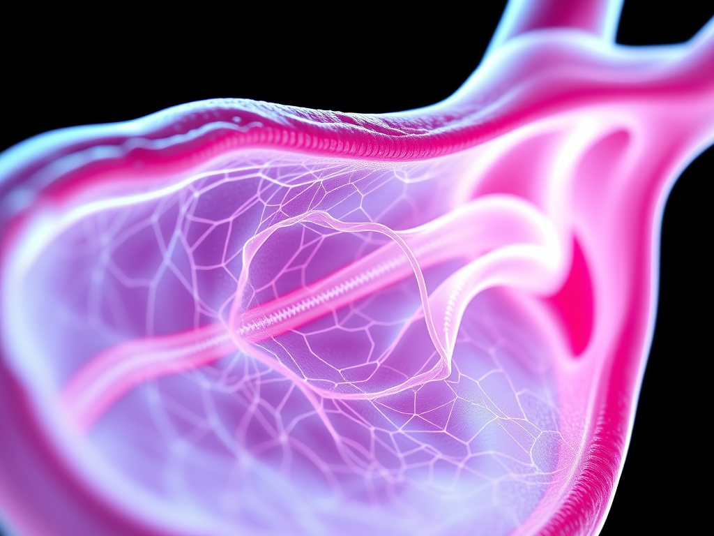
The lining of the fallopian tubes
The lining of the this tubes is a mucous membrane.
that is continuous with the mucous membrane inside the uterus. It is covered with tiny, hair-like cells called cilia that “sweep”.
the fertilized ovum down to the uterus with the currents they set up.
The lining of the this tubes is covered with delicate folds of tissue that nourish the fragile fertilized egg (Fig. 3-11).
Very small muscle fibers in the walls of this tubes. thought to be stimulated by the ovary’s release of a mature egg.
And also it create a gentle suction that draws the egg into the fimbria of the fallopian tubes.
So Tiny glands in the walls of this tubes produce nutrients that sustain the fertilized egg on its trip toward the uterus.
All adaptations work together to help ensure the implantation of a fertilized egg in the lining of the uterus.
It generally takes 3–7 days for a fertilized egg to reach the uterus.